3D Endocardial mapping
Principal investigators: Rikke Buhl and Prash Sanders
Study Director: Eva Zander Hesselkilde and Dominik Linz
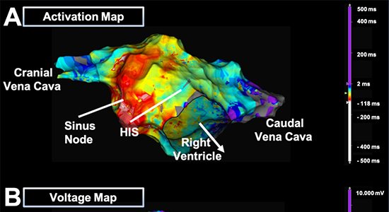
3D endocardial mapping has long been used in human medicine to characterize and treat complex arrhythmias. Especially the treatment of atrial fibrillation (AF) via ablation has dramatically evolved with the development of these mapping systems. Most of these mapping systems uses magnetic fields that requires the patients to lay completely still. In collaboration the Adelaide University, we have tested a system with reference electrodes that can be attached to the skin of the patients and therefore are less sensitive to movement. The electroanatomical cardiac mapping systems creates 3D models based on the anatomy the individual heart. The navigation system can locate the catheters and will register contact with the endocardial wall. When the catheters are moved around the heart, a 3D model of the heart appears on the screen. In addition, electrical activity can be recorded by the same catheters and displayed on the model creating both activation and voltages maps.
We have now successfully preformed 3D mapping on 4 standing horses.