
Revisiting the equine athlete’s heart & Atrioventricular conduction
Background: It is well-known that human and equine athletes develop structural changes of the heart including chamber dilation and left-sided hypertrophy. Going back in history, the large heart of racehorses has fascinated humans for decades, and the finding of the massive heart (22 pound) of the legendary racehorse Secretariat, made people wonder whether it was his enormous heart that was the reason for his exceptional speed. Later, studies involving echocardiographic examinations of the heart did in fact show a correlation between heart size and the performance on the race track. However, not only is the dimensional changes of the heart recognized, it is also accepted that human athletes develop a decrease in resting heart rate and even become bradycardic. This phenomenon has been attributed to an increase in parasympathetic activity however, this concept was recently challenged and the decrease in resting heart rate was shown to be attributed to an intrinsic remodeling of pacemaker ion channels within the sinoatrial node itself. Surprisingly, it is unclear whether racehorses also develop a decrease in resting heart rate. As it has been shown that trained horses develop larger hearts it would be reasonable to expect a lower heart rate as well. However, to date, nobody has been able to clarify this.
The Equine Cardiac Group of Copenhagen set out to scrutinize whether the horses also develop training induced decrease in resting heart rate as the human athletes do.
Methods: Resting ECG recordings were collected from 201 trained and 52 untrained Standardbred racehorses from Denmark and Sweden. A two-hour modified base-apex three-lead resting ECG was collected from all horses in the time interval from 2pm to 11pm (Figure 1). Arrhythmias were manually distinguished and counted throughout the two-hour ECG recordings. P wave duration, PR interval, QRS complex duration, QT interval and T wave duration were measured, and the P and T wave morphology noted in five to seven continuous beats (10 seconds) where the resting heart rate was at its lowest.

Results: Resting heart rate was significantly lower in the trained horses, and significantly more trained horses exhibited second-degree atrioventricular blocks (2AVB). In fact, the study revealed a general prevalence of 2AVB among horses to be 87.5 % regardless of training status when inspecting 24 hour ECG recordings, this is much higher than the current reported prevalence. Corrected QT interval was significantly shorter in trained horses. P wave duration was significantly shorter in mares compared to geldings. Stallions had significantly shorter PR intervals compared to geldings, but significantly longer QRS complex durations. Further, more trained horses exhibited monophasic T waves oppose to biphasic T waves.
Conclusion: This study is the first to reveal a decreased resting heart rate in athletic horses where a large study population and a control group were included. Further, the slowing of the AV conduction in the trained horses is compatible with the human athletic phenotype.
Atrioventricular conduction
Background: In the aforementioned study, it became clear that also the atrioventricular (AV) conduction was affected by training as more trained horses presented with second-degree AV block (2AVB). The slowing of the AV conduction is a common finding in human athletes, who also present with prolonged PR interval and first and second-degree AV block. Once again this alteration has been suggested to be due to an increase in vagal tone. This was further explored in a subgroup of horses.
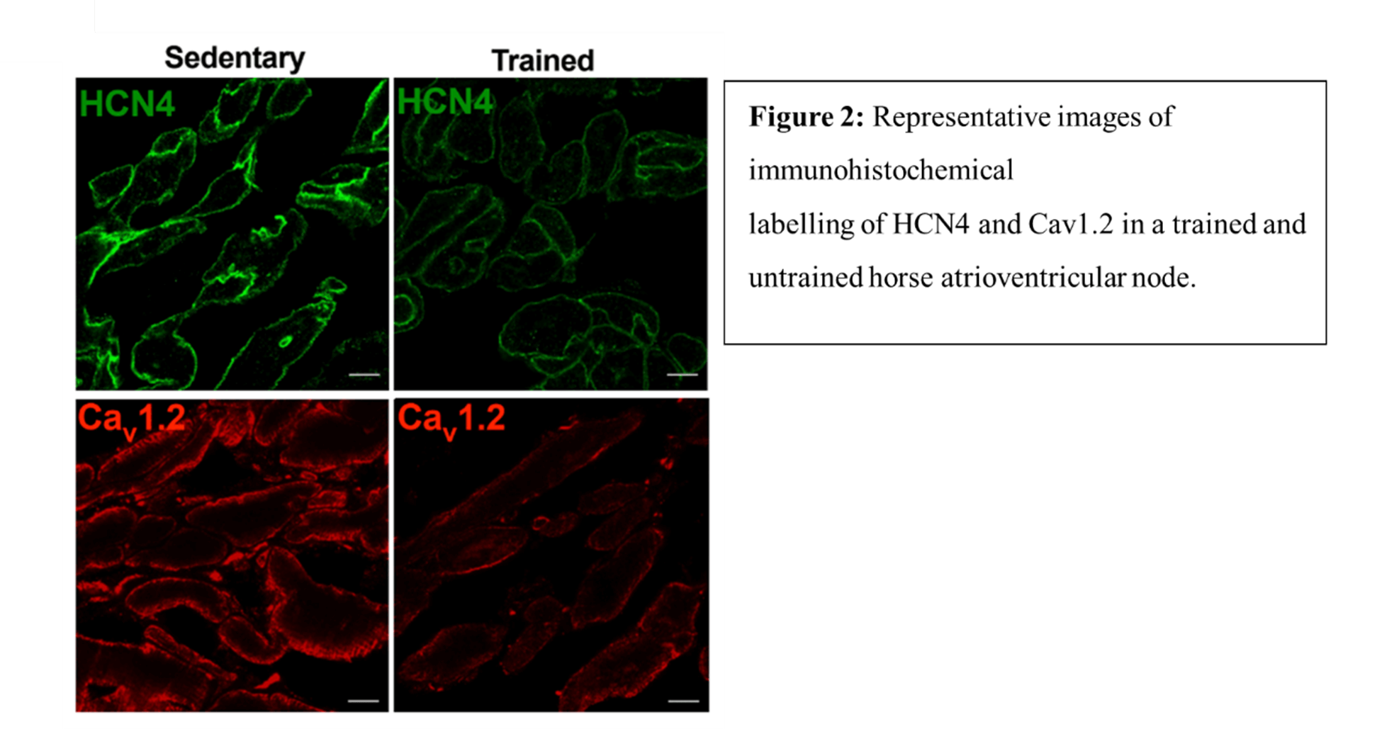
Methods: Five trained and five untrained horses underwent complete pharmacologic block of their autonomic nervous system using propranolol (0.2mg/kg) and atropine (0.04 mg/kg) IV. Assessment of the PR interval and 2AVB were conducted before and after the pharmacologic block. Further, the AV node was excised from seven trained and seven untrained horses and analysed for the expression of two key pacemaker ion channels including the hyperpolarization-activated cyclic nucleotide-gated channel 4 (HCN4) and the L-type calcium channel (CaV1.2). The expression was quantified by both immunohistochemical labelling using specific fluorescent conjugated antibodies (Figure 2) and western blot.
Results: 2AVB was completely abolished after blocking of the autonomic nervous system, however the prolonged PR interval seen in the trained horses persisted. Therefor the mechanism of the PR interval prolongation could not exclusively be due to an increase in vagal tone and the role of intrinsic remodelling of the AV node was considered for further analysis. The immunohistochemistry and western blot revealed a decrease in the expression of HCN4 and CaV1.2 proteins in the trained horses (for representative see Figure 2). A down-regulation of these two ion channels could potentially cause a delay in nodal cellular depolarisation and therefor slow down the impulse generation and conduction through the AV node. The mechanism was further explored in a mouse model confirming these findings and further revealed decreased current densities of If and Ica,L using whole cell patch clamping. Exploration of the down-stream mechanism revealed that the mRNA expression of HCN4 and CaV1.2 was under the control of miRs, and more specifically an upregulation of miR-211-5p and miR-432 proved to suppress the transcription of HCN4 and Cav1.2.
Conclusion: We conclude that intrinsic remodeling is a key mechanism underlying AV block in trained horses.
We would like to thank all the owners and trainers who have agreed to involve their horses in our studies. Without their support, none of this would have been possible.
Funding: The projects were generously funded by the Danish Council for Independent Research (DFF‐1331‐00313B) and Novo Nordisk Foundation under grant agreement number NNF18SA0034956.